DiATOME cryo AFM and DiATOME ultra AFM diamond knife.

The Diatome cryo AFM and ultra AFM knives have been developed for specimen surface investigation using Atomic Force Microscopy. Diatome ultra AFM and Diatome cryo AFM knives are specially tested to ensure that they meet the increased quality requirements of AFM investigation. They produce extremely smooth sample surfaces and guarantee the best possible structure preservation.
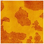 Morphology of a blend of two SBS block copolymers with different chain-architecture. AFM tapping mode, phase image, image size 3 x 3µm.
Rameshwar Adhikari, Institut fur Werkstoffwissenschaft, Martin-Luther-Universitat, Halle-Wittenberg.
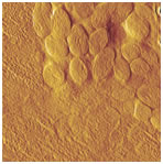 AFM amplitude image of the muscle of cat’s mite Otodectes cynotis. The contrast covers amplitude variation in the 1 – 3 nm range. Size of the whole image equals 4.6µm.
Nadejda Borisovna Matsko, Institut fur angewandte Physik, ETH Zurich.
Specifications
DiATOME cryo AFM and DiATOME ultra AFM
| Knife angle: | 35° |
| Cutting range: | 10 – 100nm |
| Available Sizes: | 2mm, 3mm |
References
P.H. Vallotton, M.M. Denn, B.A. Wood and M.B. Salmeron: Comparison of medical-grade ultrahigh molecular weight polyethylene microsctructure by AFM and TEM.
J. Biomater. Sci. Polymer Edn., Vol 6, No. 7, pp. 609-620, 1994.
N. Matsko and M. Müller: AFM of biological material embedded in epoxi resin.
Journal of Structural Biology 146, pp. 334-343, 2004.